Use of radiation to treat cancer
Radiology
Radiology is the use of radiation and radioactive substances both inside and outside the body to produce images of organs and tumours to help medical professionals identify issues and create treatment plans. Radiology is an important step in modern medical diagnosis and is crucial for locating internal cancers and organ abnormalities before treatment can be performed.
Early discovery and development
The simple x-ray (1895-96)
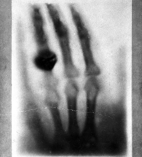
The first form of radiology ever performed was was a simple x-ray in November 1895 by German engineer and physicist Wilhelm Röntgen. An x-ray is a high energy, short wavelength electromagnetic wave capable of passing through many objects including the human body. When used in a medical context, these can produce a 2 dimensional image of internal body structures, either projected directly onto film, or using a solid-state detector where a digital image can be created. This works as more dense material or tissue, such as bones, prevent more x-rays from passing through than softer, less dense tissue like skin. The discovery of x-rays had a near instant impact on medical diagnosis, being used within a year of it's first public demonstration [1]. It should be noted this was without a proper understanding of the dangers of radiation.


The Fluoroscope (1896)
One of the first further applications of the simple x-ray was to create a real time video instead of just an image, within weeks of the demonstration of the simple xray, several such devices were created. Enrico Salvioni created the "Cryptoscope", William Francis Magie created the "Skiascope" and American inventor and businessman Thomas Edison created the "Vitascope" before coining the name "fluoroscope"[2]. This worked by placing a patient between an x-ray source and a screen coated in fluorescent metal salt, such as zinc sulphide or calcium tungstate. X-rays would pass through the body, similar to a simple x-ray but instead of leaving an image on film, the interaction between the x-ray and the fluorescent salt would produce visible yellow/green light, allowing physicians to witness the bodily functions as they happened. Edison stopped his development of this technology in the opening years of the 20th century as his assistant Clarence Dally died in 1904 of cancer induced by x-ray exposure [3]. Modern fluoroscopes are far less dangerous, using far lower dosages and are used commonly in surgeries and procedures in which objects are inserted into the body.
Mammography (~1913)
The simple x-ray could be applied to any part of the body, however certain regions of the body contain very little dense material and as such it is hard to distinguish what a cancerous tumour looks like. Mammography refers to performing x-rays on breast tissue, in 1913 a German surgeon called Albert Salomon studied 3000 mastectomies and compared it to x-ray images in order to identify what breast cancer looked like on an x-ray, however it would not become a common screening practise until 1966. A simple mammogram compresses breast tissue between 2 plates where x-rays are passed from one plate to a detector on the other, the detector can either be film or a solid-state detector to produce a digital image. In more modern times, a more advanced form of mammography called tomosynthesis can be performed where images are taken at multiple angles to produce a 3 dimensional scan of the tissue, this was first performed by researchers at Massachusetts General Hospital in 1995. It uses lower dosages of radiation than a CT scan but more than a traditional 2 dimensional scan [4].


Angiography (1927)
Blood vessels are not clear on a standard x-ray as they do not prevent the passage of radiation, as such it can be difficult to diagnose problems with blood flow. The solution to this is to inject a contrast agent (usually containing iodine or gadolinium [5]) into the blood stream which does show clearly on an x-ray allowing doctors to identify where blood is flowing, and more importantly where is is not, this is known as an angiogram. Angiography performed on the aorta may also be referred to as an aortogram. One of the first demonstrations of angiography was performed by Egas Moniz in 1927 and up until the 1970s remained the only way to produce an image of blood vessels [6]. This can be used to identify conditions such as blood clots and brain aneurysms.
Short Quiz on early discovery
Modern developments and technology
Computerised Tomography (1971)
A CT scanner makes use of a motorised x-ray source that rotates around a patient with a detector on the far side. After a full rotation this will scan a cross section of the patient between 1 and 10mm thick, the bed then moves further into the scanner and another cross section is scanned. The process repeats until the entire target region has been covered, the images can be viewed individually or they can be compiled into a 3 dimensional image of the whole region. This can provide far more in depth analysis than a simple x-ray and can be used to identify (but is not limited to) tumours, blood clots, heart abnormalities and complex bone fractures. The first use of a CT scanner on a live patient was in October 1971 and within the next few years found it's way into hospitals [7]. CT scans are very common today, with NHS England performing over six million (~6,647,255) CT scans in the period covering January 2022 to January 2023 [8].


Nuclear Medicine (~1960s-1970s)
Nuclear medicine, in contrast to the previous methods described makes use of internal radiotracers instead of external radiation sources. The two main types are Positron Emission Tomography (PET) and Single Photon Emission Computerised Tomography (SPECT).
A PET scan uses a rapidly decaying compound similar to natural glucose, commonly fluorodeoxyglucose (FDG). As cancers are rapidly replicating, they drain large amounts of the body's glucose and as such, upon entering the bloodstream, the radiotracer is absorbed into the tumour which can be viewed using a PET camera. On the contrary, when screening for conditions like dementia, the radiotracer will not be present in the decaying region, making it easy to identify. This can be used in conjunction with CT and MRI scanners [9].
A SPECT scan injects one of a variety of gamma emitting radiotracers into the blood stream; including, but not limited to: iodine-123, technetium-99m, xenon-133, thallium-201 and fluorine-18. Gamma radiation is another form of electromagnetic wave that has more energy than an x-ray and can pass trough most substances with ease, conveniently it can be detected using a CT scanner, meaning both cross sections and a 3 dimensional image can be produced. This is primarily used to show how blood flows through a body and can highlight blockages and organ abnormalities [10].
Magnetic Resonance Imaging (1977)
MRI is one of the newest and arguably most complex forms of radiology with the benefit of not using ionising radiation. The scanner employs large magnets and by extension a powerful magnetic field to align all the protons (a positively charged sub-atomic particle present in every atom) in the body, radio-waves (a low energy electromagnetic wave) are then pulsed through the patient which force the protons against the pull of the magnetic field. When the radio-waves are removed, the protons release energy as they fall back into alignment with the magnetic field and this energy is detected by MRI sensors. As chemical structure and environment effect the amount of energy released, radiologists can easily distinguish between parts of the body [11].
The first use of an MRI scanner on a live patient was in 1977 and it's development can be attributed to the work of American physicist and Nobel Prize winner Isidor Isaac Rabi [12].
